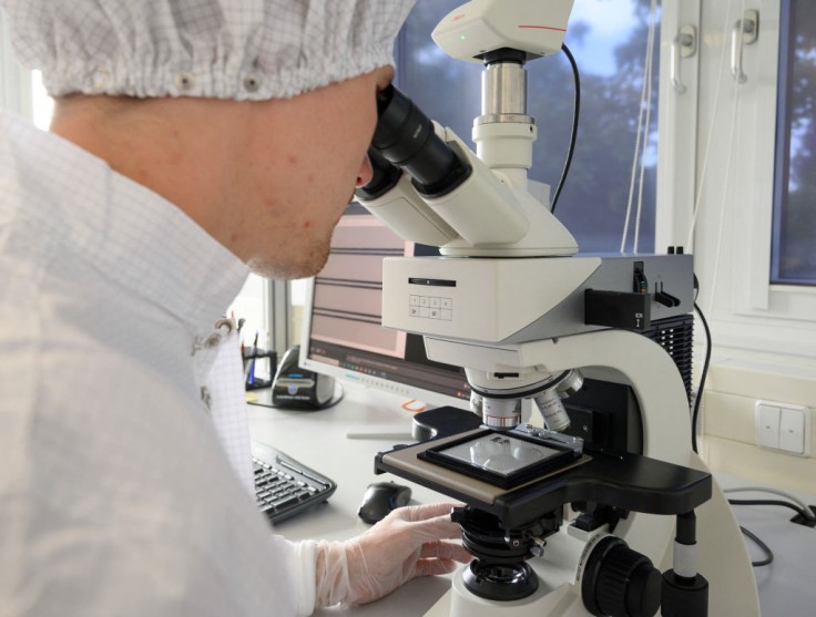
Through a special X-ray microscope, an interdisciplinary research group from the University of Gottingen and Hannover Medical School (MHH) discovered a significant abnormality in the cardiac muscle tissue of people who died with COVID-19.
Special X-Ray Microscope
For a long time, lung tissue damage has been the focus of the researchers, and it has now been comprehensively examined.
"The current study underpins the involvement of the heart in COVID-19 at the microscopic level for the first time by imaging and analyzing the affected tissue in the three dimensions," the researchers explained, per Medical Express.
Moreover, the researchers used synchrotron radiation, which is very intense X-ray radiation, to photograph the tissue architecture at a high resolution and show it three-dimensionally.
The researchers did this with the help of a specialized X-ray microscope built by the University of Gottingen and operated at the German Electron Synchrotron DESY in Hamburg.
When they looked at the impacts of the severe type of COVID-19 illness on the capillaries (small blood vessels) in cardiac muscle tissue, they saw evident abnormalities.
As compared to a healthy heart, an X-ray image of tissues damaged by severe illness showed a network of splits, branches and loops that had been unevenly modified by the formation and splitting of new arteries.
These changes in the tissue are the first direct visible evidence of one of the main causes of lung injury in COVID-19. In scientific terms, it is considered as a kind of "intussusceptive angiogenes," meaning new vessel development.
How Does the Special X-Ray Microscope Detect COVID-19 Damage to the Heart?
To show the capillary network, machine learning approaches were used to identify the vessels in the three-dimensional volume. Initially, researchers had to carefully label the visual data by hand.
The first author of the work from the University of Gottingen Marius Reichardt stated that "to speed up image processing, we therefore also automatically broke the tissue architecture down into its local symmetrical features and then compared them."
Moreover, Professor Tim Salditt of the University of Gottingen and Professor Danny Jonigk of the MHH explained that "the parameters obtained from this then showed a completely different quality compared to healthy tissue, or even to diseases such as severe influenza or common myocarditis."
In contrast to vascular architecture, the necessary data quality could be obtained in the University of Gottingen's laboratory utilizing a tiny X-ray source. This implies that it could theoretically be used in any clinic to assist pathologists with regular diagnoses.
The researchers hope to expand their technique of turning typical tissue patterns into abstract mathematical values in the future in order to build automated diagnostic tools. They look to achieve this by further refining laboratory X-ray imaging and testing it with synchrotron radiation data.
Another Special X-Ray Technique Shows Vascular Damage in COVID-19 Lungs
Aside from the mentioned Special X-Ray Microscope, the new X-ray technology operates similarly to a hospital computed tomography (CT) scan, but its resolution is a hundred times greater, per Medica Magazine.
Moreover, Hierarchical Phase Contrast Tomography (HiP-CT) is a revolutionary method that can capture the tiniest vessels with a diameter of five micrometers or roughly one-hundredth of a hair's thickness. HiP-CT allows researchers to see into the lungs' depths and visualize even the tiniest structures, down to individual cells.


